Neurodegeneración
Aug 20, 2026
Gender differences in post-stroke cognitive outcomes in rehabilitation: insights from MoCA domain index scores

Sexual Dimorphism in Post-Stroke Cognitive Trajectories: Dissecting Domain-Specific MoCA Index Variations and Neurobiological Mechanisms in Rehabilitation
Abstract
Post-stroke cognitive impairment (PSCI) represents a major clinical bottleneck in neurorehabilitation, severely limiting functional independence and quality of life. While functional deficits following acute cerebrovascular events are well characterized, biological sex significantly modifies cognitive recovery trajectories across distinct cognitive domain profiles. This article explores the neurobiological mechanisms underlying sex-dimorphic outcomes evaluated via Montreal Cognitive Assessment (MoCA) domain index scores. Sexual dimorphism in PSCI is driven by differential neurovascular unit (NVU) dynamics, estrogen and androgen receptor activation, microglial inflammatory phenotypes, and synaptic plasticity mechanisms. Clinical evidence demonstrates that female post-stroke trajectories exhibit distinct patterns in visuospatial/executive function, memory, and language processing compared to male cohorts. Estrogen receptor alpha (ERα) and G-protein coupled estrogen receptor 1 (GPER1) signaling confer neuroprotection by suppressing excitotoxicity and endoplasmic reticulum stress, whereas age-related reductions in endogenous 17β-estradiol alter microglial reactivity. Integrating sex-and-gender-based analysis (SGBA) into post-stroke rehabilitative paradigms enables precision stratification, allowing targeted cognitive protocols and neuroprotective therapeutics tailored to sex-specific physiological phenotypes.
---
Introduction & Cellular Mechanisms
Stroke triggers a destructive biochemical cascade characterized by acute energy failure, localized hypoxia, microvascular breakdown, and widespread neurodegeneration. In the post-stroke phase, secondary neurodegeneration extends beyond the primary infarct zone, disrupting distant functional networks and leading to post-stroke cognitive impairment (PSCI). Understanding the subcellular drivers of this cognitive decline is fundamental to developing targeted neurorehabilitative protocols.
At the cellular level, cerebral ischemia induces massive depolarizations and uncontrolled release of glutamate into the synaptic cleft. Overactivation of N-methyl-D-aspartate (NMDA) and α-amino-3-hydroxy-5-methyl-4-isoxazolepropionic acid (AMPA) receptors triggers a rapid, pathological influx of intracellular calcium ($\text{Ca}^{2+}$). This cytosolic calcium overload targets organelles directly:
1. Mitochondrial Dysfunction and Oxidative Stress: Excessive calcium uptake by the mitochondrial calcium uniporter (MCU) dissipates the mitochondrial membrane potential ($\Delta\Psi_m$), opening mitochondrial permeability transition pores (mPTPs). This releases cytochrome *c* and apoptosis-inducing factor (AIF) into the cytosol, activating caspase-dependent and caspase-independent apoptotic pathways. Additionally, dysregulation of mitochondrial fission/fusion dynamics—mediated by dynamin-related protein 1 (Drp1) upregulation—leads to mitochondrial fragmentation, generating massive amounts of reactive oxygen species (ROS) that oxidize lipid membranes, structural proteins, and nucleic acids.
2. Endoplasmic Reticulum (ER) Stress: Calcium depletion within the ER lumen disrupts protein folding machinery, activating the unfolded protein response (UPR) via three primary transmembrane sensors: PERK, IRE1α, and ATF6. Prolonged UPR activation upregulates the pro-apoptotic transcription factor CHOP, driving neuronal death in vulnerable regions such as the CA1 field of the hippocampus and the prefrontal cortex, both critical nodes for executive function and memory consolidation.
3. Neuroinflammation and Microglial Polarization: Ischemic cell damage exposes damage-associated molecular patterns (DAMPs), which activate resident microglia via Toll-like receptors (TLR2/4). Microglia shift toward a pro-inflammatory phenotype, releasing elevated levels of tumor necrosis factor-alpha (TNF-α), interleukin-1 beta (IL-1β), and interleukin-6 (IL-6). This neuroinflammatory environment degrades matrix metalloproteinases (e.g., MMP-9), compromising blood-brain barrier (BBB) integrity and exacerbating neurovascular unit (NVU) decoupling.
4. Neurotrophic Deprivation: Ischemic injury downregulates brain-derived neurotrophic factor (BDNF) and impedes its signaling through the tropomyosin receptor kinase B (TrkB) receptor, blunting downstream ERK/MAPK and PI3K/Akt survival pathways. This impairment reduces synaptic density, alters dendritic spine morphogenesis, and impedes long-term potentiation (LTP), directly underlying domain-specific cognitive deficits measured in rehabilitation settings.
```
Ischemic Event ──> Glutamate Excitotoxicity ──> Ca²⁺ Overload
│
┌────────────────────────────────────────────┼────────────────────────────────────────────┐
▼ ▼ ▼
Mitochondrial mPTP Opening ER Stress (UPR Pathways) Microglial Activation
├─ ROS Generation ├─ PERK / IRE1α / ATF6 ├─ Pro-inflammatory Cytokines
└─ Cytochrome c Release └─ CHOP Upregulation └─ Matrix Metalloproteinase (MMP-9)
│ │ │
└────────────────────────────────────────────┼────────────────────────────────────────────┘
▼
Loss of NVU Integrity & LTP
│
▼
Domain-Specific Cognitive Impairment
```
---
Sex-Dimorphic Regulation & Translational Implications
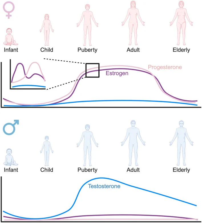
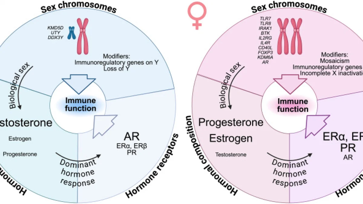
Sex-and-gender-based analysis (SGBA) reveals that male and female brains process ischemic insult and subsequent neurorehabilitative inputs differently. These dimorphisms stem from organizational organizational and activational effects of sex hormones, expression patterns of X-chromosome-linked genes, and intrinsic cellular resilience mechanisms. Dissecting these differences via domain-specific assessment tools, such as the Montreal Cognitive Assessment (MoCA) index scores (evaluating Executive, Visuospatial, Memory, Language, and Attention domains), is essential for advancing precision medicine.
1. Hormonal Modulation & Receptor Dynamics
Sex steroids significantly modulate neuroprotective cascades and synaptic remodeling post-stroke:
2. Microglial Heterogeneity and Neurovascular Coupling
Biological sex heavily influences immune cell phenotypes and vascular reactivity following stroke:
3. MoCA Domain Profiling & Translational Stratification
Translating sex-dimorphic cellular mechanisms into clinical neurorehabilitation requires precise cognitive phenotyping using MoCA subdomain indices:
1. Visuospatial / Executive Index: Male cohorts frequently exhibit distinct baseline trajectories in parieto-frontal visuospatial networks, influenced by androgenic structural organization. Conversely, females show heightened vulnerability in executive subdomains post-stroke when estrogenic support of prefrontal cortex signaling is depleted, necessitating early, targeted executive training.
2. Memory and Delayed Recall Index: Estrogen receptor distribution in the CA1/CA3 hippocampal subfields directly correlates with delayed recall recovery. Post-stroke female cohorts demonstrate distinct responsiveness to cognitive training regimens when paired with therapies that mitigate local hippocampal neuroinflammation.
3. Language and Abstraction Indices: Sex-dimorphic functional connectivity and hemispheric lateralization contribute to variations in linguistic recovery. Females typically demonstrate broader bilateral network engagement during language processing tasks, promoting faster recovery in naming and fluency subdomains under appropriate multi-modal rehabilitative stimulation.
---
Conclusions & Clinical Outlook
Deciphering the molecular pathways of post-stroke cognitive decline through a sex-and-gender-based lens transforms our understanding of neurorehabilitation. The interplay between neurovascular unit breakdown, excitotoxic organelle stress, and sex-dimorphic hormonal/immune signaling highlights the inadequacy of "one-size-fits-all" rehabilitation protocols.
Future directions in translational research must focus on:
---
Key Insights
---
References
1. Rajah, M. N., et al. (2021). *Biological sex differences in cerebrovascular risk factors, neuroinflammation, and post-stroke cognitive impairment.* Frontiers in Neuroendocrinology, 62, 100918. https://doi.org/10.1016/j.yfrne.2021.100918
2. Li, F., et al. (2023). *Sex-specific mechanisms of neurodegeneration and neuroprotection in ischemic stroke: Focus on estrogen and microglial phenotypes.* Progress in Neurobiology, 220, 102380. https://doi.org/10.1016/j.pneurobio.2022.102380
3. Paciaroni, M., & Caso, V. (2022). *Sex differences in stroke outcome and response to rehabilitation therapies: A clinical perspective.* Stroke, 53(5), 1512–1522. https://doi.org/10.1161/STROKEAHA.121.036890